Abstract
Percutaneous closure of the left atrial appendage (LAA) is an alternative treatment for stroke prevention in patients with atrial fibrillation who cannot tolerate oral anticoagulation. The conventional method involves transseptal puncture to access and seal the LAA. However, recent studies have investigated the possibility of performing LAA closure through a patent foramen ovale (PFO) tunnel. This case aims to illustrate the simultaneous closure of both the LAA and PFO using the PFO tunnel as the access route.
Keywords: Interventional cardiology, left atrial appendage, patent foramen ovale, percutaneous closure
INTRODUCTION
Atrial fibrillation (AF) is the most common arrhythmia, with its prevalence increasing with age. Stroke is a frequent complication of non-valvular AF (NVAF). Percutaneous left atrial appendage (LAA) closure is a key treatment for stroke prevention in patients with NVAF, particularly those who cannot tolerate oral anticoagulation. Both the LAA in patients with AF and a patent foramen ovale (PFO) or atrial septal defect (ASD) are major sources of cardioembolic stroke. Combining LAA closure with PFO/ASD closure may offer an optimal approach to stroke prevention. This report describes a case of simultaneous percutaneous closure of a PFO and LAA.
CASE REPORT
A 76-year-old woman with permanent AF, hypertension, diabetes mellitus, and dyslipidemia was referred for transesophageal echocardiography (TOE) and LAA closure. One month earlier, she had been hospitalized for massive gastrointestinal bleeding while on warfarin, requiring a blood transfusion of approximately 14 units. Her CHA2DS2-VASc and HAS-BLED scores were both 5. She was switched to apixaban at discharge but subsequently experienced an ischemic stroke while on medication.
Electrocardiography showed AF with a mean heart rate of 90 bpm. Chest radiography was remarkable, and there was no significant family history. Blood tests revealed mildly elevated glucose and cholesterol levels, along with an increased D-dimer level. Physical examination showed mild swelling in the left leg, and Doppler ultrasonography confirmed deep vein thrombosis in the same leg. Preprocedural TOE identified a small shunt in the interatrial septum, consistent with a PFO (, ). A bubble study demonstrated a significant right-to-left shunt, and the LAA was visualized with no thrombus (, ). Given her medical history and comorbidities, simultaneous PFO and LAA closure was selected to eliminate the need for oral anticoagulation.
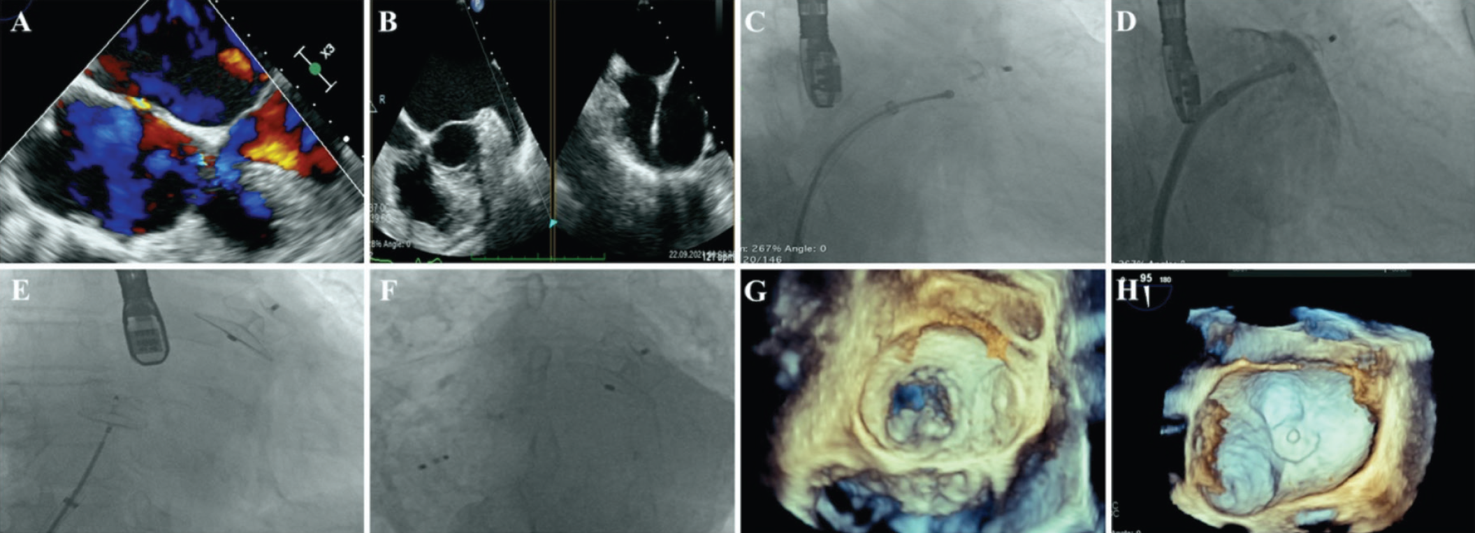
Intravenous proton pump inhibitor therapy was initiated, and oral anticoagulation was replaced with subcutaneous low-molecular-weight heparin. The procedure was performed under general anesthesia using right femoral vein access, with fluoroscopic and 2D/3D TOE guidance. A 25-mm Amplatzer Amulet LAA occluder and a 25-mm Amplatzer PFO occluder were selected. Transseptal access was achieved using an SL 1 sheath and Inoue wire, followed by the insertion of a 12 Fr delivery sheath.
The LAA occlusion device was carefully advanced, and its position was confirmed using fluoroscopy and TOE (, ). Deployment was adjusted to ensure optimal placement, with contrast imaging used to verify device position and assess for leakage (, ). The PFO occluder was implanted using the standard technique (, ). Device positioning was evaluated in the left anterior oblique view (). A follow-up TOE performed 45 days later showed no device-related complications, with both the PFO or LAA occluders in stable positions ( and ).
DISCUSSION
The PFO was positioned anterosuperiorly, while the LAA was located anterolaterally. Transseptal access via the PFO can be challenging due to the cranioanterior orientation of the LAA. Proper alignment may require external rotation and the assistance of two operators. Studies have reported high success rates for device implantation, with major complication rates similar to those of standard techniques. Long-term outcomes of transseptal access through a PFO or ASD have demonstrated comparable efficacy in stroke prevention and similar safety profiles. Simultaneous PFO/ASD closure did not prolong fluoroscopy time but required a higher contrast volume. Additionally, this approach was associated with a lower incidence of stroke and transient ischemic attacks compared to other methods.
CONCLUSION
Transseptal access via a PFO or ASD allows entry into the left atrium while reducing the risk of complications associated with transseptal puncture, such as perforation of the left atrial free wall or aortic root. Compared to transseptal puncture, utilizing a PFO or ASD for left atrial access is a viable and safe alternative when using Amplatzer systems for LAA closure. Additionally, performing simultaneous PFO or ASD closure may offer further protection against systemic embolization without increasing procedural risk.
Video 1. Transesophageal echocardiogram visualization of the patent foramen ovale shunt
Video 2. Confirmation of the absence of thrombus in the left atrial appendage
Video 3. Implantation of the Amulet device
Video 4. Verification of no contrast leakage upon injection
Video 5. Implantation of the patent foramen ovale occlusion device
Footnotes
Informed Consent: Written consent was obtained from the patient and their relatives for the inclusion of case details and personal information.
Authorship Contributions: Surgical and Medical Practices: Z.E.G., R.Z., Concept: R.Z., Design: R.Z., Data Collection or Processing: Z.E.G., Analysis or Interpretation: R.Z., Literature Search: Z.E.G., Writing: Z.E.G.
Conflict of Interest: No conflict of interest was declared by the authors.
Financial Disclosure: The authors declared that this study received no financial support.
References
Tzikas A, Shakir S, Gafoor S, et al. Left atrial appendage occlusion for stroke prevention in atrial fibrillation: multicentre experience with the AMPLATZER Cardiac Plug. EuroIntervention. 2016;11:1170-1179.
Koermendy D, Nietlispach F, Shakir S, et al. Amplatzer left atrial appendage occlusion through a patent foramen ovale. Catheter Cardiovasc Interv. 2014;84:1190-1196.
Kleinecke C, Fuerholz M, Buffle E, et al. Transseptal puncture versus patent foramen ovale or atrial septal defect access for left atrial appendage closure. EuroIntervention. 2020;16:e173-e180.
Supplementary Materials
Video 1. Transesophageal echocardiogram visualization of the patent foramen ovale shunt
DownloadVideo 2. Confirmation of the absence of thrombus in the left atrial appendage
DownloadVideo 3. Implantation of the Amulet device
DownloadVideo 4. Verification of no contrast leakage upon injection
DownloadVideo 5. Implantation of the patent foramen ovale occlusion device
Download