INTRODUCTION
A 43-year-old male patient with a known history of hypertension presented to the emergency department of an external center with chest pain. He was referred to our hospital for coronary angiography with a diagnosis of acute lateral myocardial infarction (MI), based on ST elevation in leads D1, AVL, and V5-6, and reciprocal ST depression in leads D2, D3, and arteriovenous fistulas (AVF) on his electrocardiography (ECG) (). Upon arrival at our clinic, the patient was promptly taken to the catheterization laboratory. He reported relief of chest pain, and coronary angiography revealed no lesion in the right coronary artery. No total occlusion was observed in the left anterior descending (LAD) or circumflex (CX) arteries during left coronary system imaging. However, a lesion in the proximal LAD was notable (). The patient reported that his chest pain persisted, although less intense. The ECG also showed persistent ST elevation.
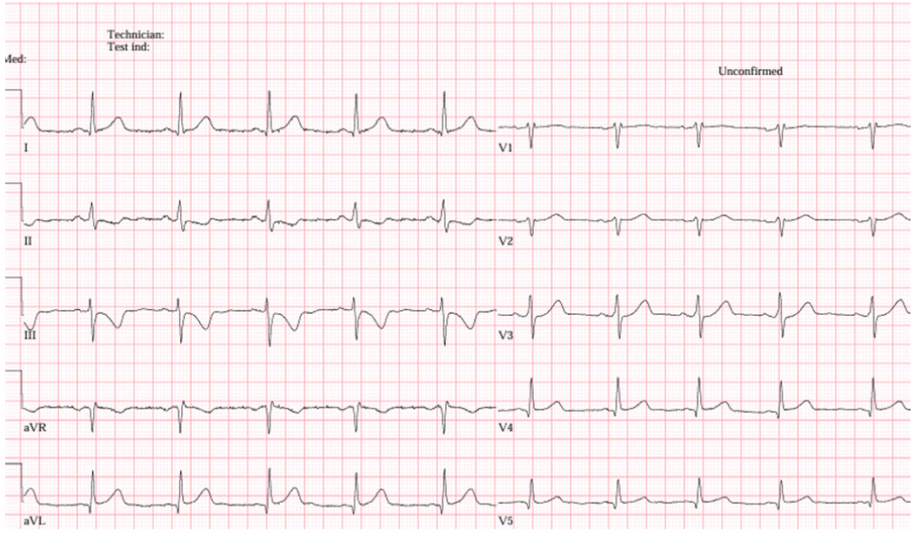
ECG: Electrocardiogram
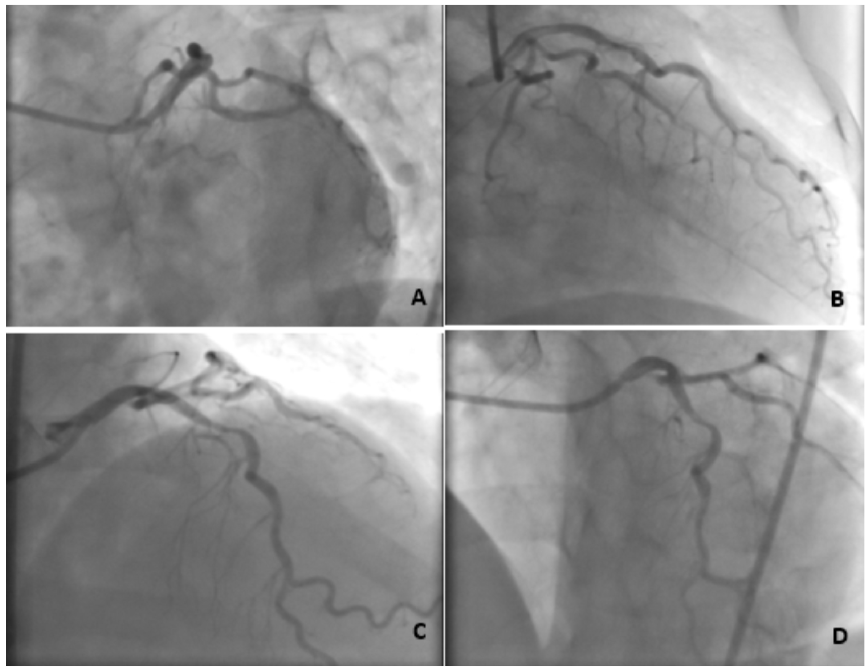
LAD: Left anterior descending
Therefore, based on the persistent ischemic findings on ECG, the LAD was wired, and percutaneous transcatheter coronary angioplasty (PTCA) was performed at the proximal lesion. The thrombus at the LAD lesion site had occluded the ostium of the diagonal artery (DG), and PTCA at this site restored flow in the DG. Subsequently, a 3.0 × 26 mm drug-eluting stent with optimal apposition was implanted at the culprit lesion in the LAD. Proximal optimization was performed with a 3.5 × 12 mm non-compliant balloon. LAD-DG kissing PTCA was then carried out, and the LAD stent was further optimized. Control images confirmed restoration of distal flow in both the LAD and the DG (). The patient’s chest pain resolved, and the procedure was successfully completed.
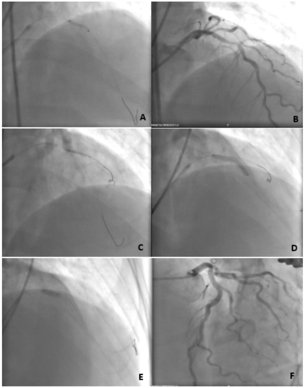
PTCA: Percutaneous transcatheter coronary angioplasty, LAD: Left anterior descending, DG: Diagonal artery
Lateral MI is diagnosed on ECG by ST elevation in leads D1, AVL, and V5-6, with reciprocal ST depression in leads D2, D3, and AVF. Early revascularization significantly improves clinical outcomes. Moreover, percutaneous intervention has shown better results than fibrinolytic therapy. Hreybe et al. reported an increased risk of ventricular fibrillation in patients presenting with anterior and lateral MI. Lateral MI may result from obstruction of the obtuse marginal artery, DG, or ramus intermedius. In our case, although the DG—completely occluded—was not visible on coronary angiography, it became apparent after LAD PTCA, guided by ECG findings.
In conclusion, in patients presenting with lateral MI who have a high risk of ventricular arrhythmia, if relatively smaller vessels or side branch disease cannot be detected on coronary angiography, ECG findings should be relied upon. The ramus intermedius should be sought, and if a lesion is identified in the LAD or CX, PTCA should be performed in that region to reveal a possible side branch lesion responsible for the ischemia.
Footnotes
Informed Consent: Written informed consent was obtained from the patient.
Presented in: The study was previously presented as a paper at the 1st Interventional Cardiology Congress 2025.
Authorship Contributions: Surgical and Medial Practices: M.A.Ç., F.K., E.Ç., G.A., Concept: M.A.Ç., F.K., E.Ç., G.A., Design: M.A.Ç., F.K., E.Ç., G.A., Data Collection or Processing: M.A.Ç., F.K., E.Ç., G.A., Analysis or Interpretation: M.A.Ç., F.K., E.Ç., G.A., Literature Search: M.A.Ç., Writing: M.A.Ç., E.Ç.
Conflict of Interest: No conflict of interest was declared by the authors.
Financial Disclosure: The authors declared that this study received no financial support
References
Anderson JL, Karagounis LA, Califf RM. Metaanalysis of five reported studies on the relation of early coronary patency grades with mortality and outcomes after acute myocardial infarction. Am J Cardiol. 1996;78:1-8.
Hreybe H, Saba S. Location of acute myocardial infarction and associated arrhythmias and outcome. Clin Cardiol. 2009;32:274-277.
Thrane PG, Kristensen SD, Olesen KKW, et al. 16-year follow-up of the Danish acute myocardial infarction 2 (DANAMI-2) trial: primary percutaneous coronary intervention vs. fibrinolysis in ST-segment elevation myocardial infarction. Eur Heart J. 2020;41:847-854.