Abstract
Coronary artery fistulas (CAFs) are abnormal communications between the coronary arteries and the cardiac chambers or great vessels. Although most patients remain asymptomatic, symptomatic cases may present with dyspnea, angina, heart failure, myocardial ischemia, or myocardial infarction. We report the case of a 47-year-old woman who presented with type 2 myocardial infarction secondary to coronary steal caused by coronary-to-pulmonary artery fistulas arising from all three major coronary arteries in the presence of non-obstructive coronary artery disease. This case highlights a rare presentation of CAF associated with type 2 myocardial infarction.
Keywords: Coronary artery fistulas, type 2 myocardial infarction, myocardial ischemia
INTRODUCTION
Coronary artery fistulas (CAFs) are rare congenital or acquired coronary artery anomalies characterized by abnormal communications between a coronary artery and a cardiac chamber or great vessel. They are identified in approximately 0.1-0.2% of patients undergoing invasive coronary angiography (CAG) and are increasingly detected with the widespread use of coronary computed tomography (CT) angiography.- They most commonly originate from the right coronary artery or the left anterior descending (LAD) artery and typically drain into the right-sided cardiac chambers or the pulmonary artery. Although most patients remain asymptomatic, symptomatic cases may present with dyspnea, angina, heart failure, myocardial ischemia, or myocardial infarction., We present the case of a patient with type 2 myocardial infarction caused by the coronary steal phenomenon secondary to coronary-to-pulmonary artery fistulas arising from all three major coronary arteries in the setting of non-obstructive coronary artery disease. To our knowledge, the presentation of type 2 myocardial infarction in patients with coronary-to-pulmonary artery fistulas involving all three major coronary arteries is exceedingly rare.
CASE REPORT
A 47-year-old woman with a history of hypertension and diabetes mellitus presented to the emergency department with typical chest pain of 3 hours’ duration. She also reported exertional angina during the preceding 3 months. On admission, she was hemodynamically stable, and the physical examination was unremarkable. The initial evaluation included a 12-lead electrocardiogram (ECG), laboratory testing with cardiac biomarkers, chest radiography, and transthoracic echocardiography. The admission ECG demonstrated mild horizontal ST-segment depression in leads V1-V6, I, and aVL. High-sensitivity cardiac troponin T was elevated at 157 ng/L, whereas routine laboratory test results, including the complete blood count, were within normal limits. D-dimer levels were also within the normal range. Transthoracic echocardiography revealed preserved left ventricular systolic function without regional wall motion abnormalities. No significant valvular disease, right ventricular dilation, pericardial effusion, or other structural congenital cardiac abnormalities were identified. CAG demonstrated non-obstructive stenosis of the mid-LAD artery. In addition, coronary-to-pulmonary artery fistulas originating from all three major coronary arteries were identified (). Selective balloon occlusion of the left circumflex artery (LCx) markedly reduced shunt flow into the pulmonary artery, confirming its contribution to the fistulous circulation. Persistent opacification of the pulmonary artery via the LAD artery indicated additional fistulous communications (). Based on the clinical presentation, elevated cardiac biomarkers, ischemic ECG changes, and the absence of obstructive coronary artery disease, the patient was diagnosed with type 2 myocardial infarction secondary to the coronary steal phenomenon caused by multiple coronary-to-pulmonary artery fistulas. Percutaneous transcatheter closure of the fistulas using coil embolization was recommended; however, the patient declined the procedure. Therefore, conservative medical management was continued, and she was discharged on appropriate medical therapy with scheduled outpatient follow-up.
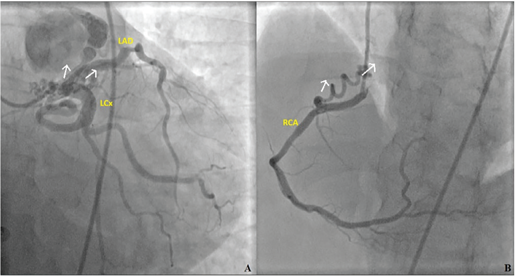
LAD: Left anterior descending artery, RCA: Right coronary artery
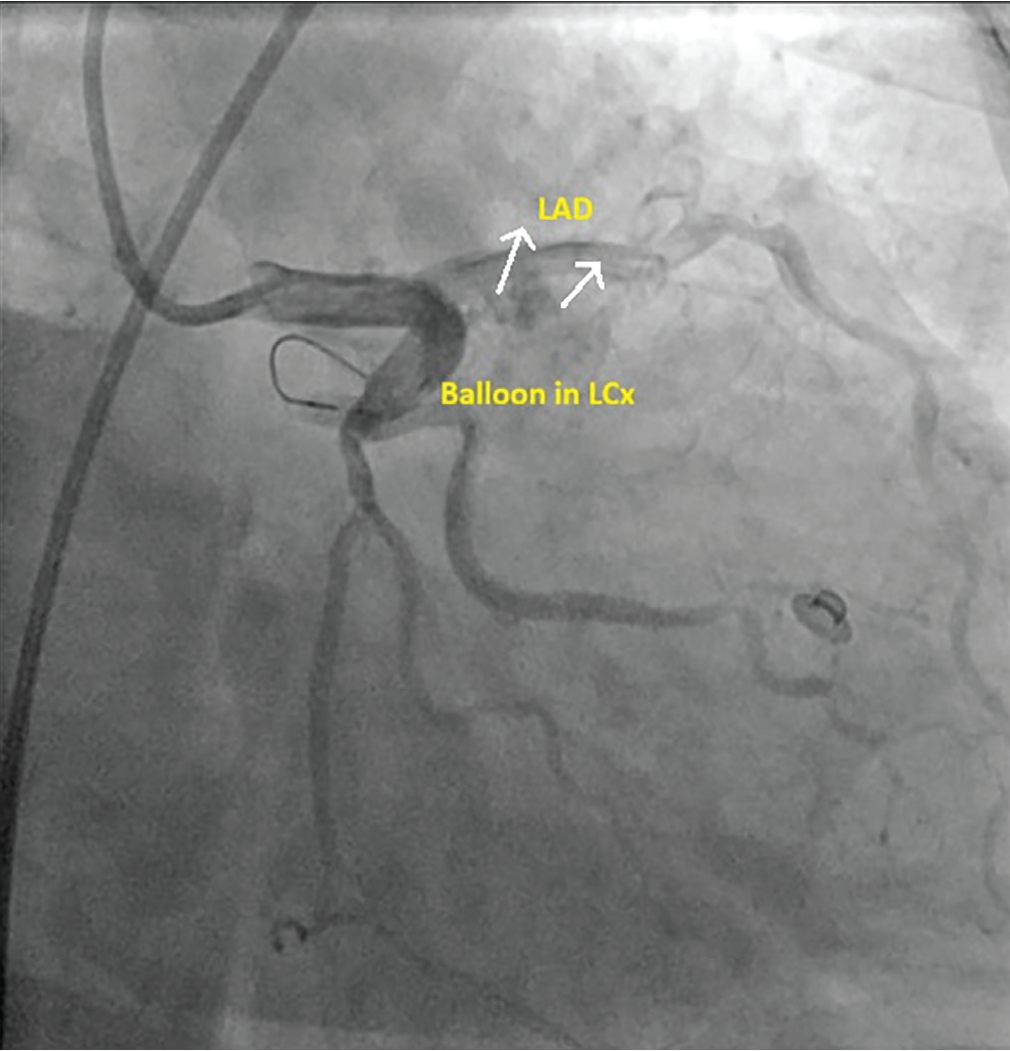
DISCUSSION
CAFs are rare coronary anomalies, with a reported prevalence of 0.2-0.6% among patients undergoing CAG. Coronary-to-pulmonary artery fistulas are the most common subtype, whereas fistulas arising from all three major coronary arteries are exceedingly uncommon. In the present case, coronary-to-pulmonary artery fistulas originated from all three major coronary arteries in the presence of non-obstructive coronary artery disease and presented clinically as type 2 myocardial infarction.
Myocardial infarction in patients with CAFs may result from several mechanisms, including flow-mediated vascular remodeling, thrombosis, coronary dissection, external coronary compression, and abnormalities in coronary perfusion pressure. However, the most widely accepted mechanism of myocardial ischemia in the absence of obstructive coronary artery disease is the coronary steal phenomenon, in which blood preferentially flows through the low-resistance fistulous tract, thereby reducing distal myocardial perfusion. According to the Fourth Universal Definition of myocardial infarction, type 2 myocardial infarction results from an imbalance between myocardial oxygen supply and demand unrelated to acute atherothrombotic plaque disruption. In our patient, the presence of typical ischemic chest pain, elevated high-sensitivity cardiac troponin T levels, and ischemic ST-segment depression fulfilled the diagnostic criteria for acute myocardial infarction. CAG demonstrated only non-obstructive coronary artery disease, with no evidence of plaque rupture or coronary thrombosis, suggesting that myocardial ischemia was most likely attributable to coronary steal through multiple coronary-to-pulmonary artery fistulas. Therefore, we considered this presentation to be consistent with type 2 myocardial infarction. In patients presenting with myocardial infarction and non-obstructive coronary artery disease, alternative mechanisms, such as myocardial infarction with non-obstructive coronary disease, coronary vasospasm, coronary embolism, and coronary microvascular dysfunction, should be considered. In the present case, however, CAG revealed multiple coronary-to-pulmonary artery fistulas without evidence of plaque rupture, coronary thrombosis, or coronary embolism. Moreover, the patient had a 3-month history of exertional angina preceding the acute presentation, a clinical pattern consistent with myocardial ischemia caused by the coronary steal phenomenon. Taken together with the absence of angiographic findings supporting alternative epicardial coronary mechanisms, coronary steal was considered the most plausible explanation for the patient’s type 2 myocardial infarction.
Previous reports have described myocardial infarction in patients with CAFs; however, the underlying mechanisms differ substantially from those observed in our case. Roule et al. reported myocardial infarction in a patient with coronary-to-pulmonary artery fistulas involving all three major coronary arteries, but significant obstructive coronary artery disease was also present. Similarly, Smettei and Abazid described acute myocardial infarction secondary to thrombosis within a CAF extending into the LCx. In contrast, our patient had non-obstructive coronary artery disease without angiographic evidence of acute plaque rupture or coronary thrombosis. Therefore, coronary steal caused by multiple coronary-to-pulmonary artery fistulas was considered the most likely mechanism of myocardial ischemia, fulfilling the diagnostic criteria for type 2 myocardial infarction.
Coronary CT angiography was not performed in this case. Although invasive CAG adequately demonstrated the coronary anatomy and fistulous tracts, CT angiography could have provided additional anatomical information regarding the course and spatial relationships of the fistulous connections.
Current evidence supports the closure of symptomatic CAFs, particularly in patients with myocardial ischemia, heart failure, significant left-to-right shunting, or other fistula-related complications. Depending on the anatomical characteristics of the fistula, closure can be achieved surgically or by transcatheter techniques. Owing to its minimally invasive nature and favorable outcomes, transcatheter closure has become the preferred therapeutic approach for anatomically suitable patients. In our patient, transcatheter coil embolization was recommended because of symptomatic myocardial ischemia; however, the patient declined the procedure and was therefore managed conservatively with close clinical follow-up.
CONCLUSION
This case expands the clinical spectrum of coronary-to-pulmonary artery fistulas by demonstrating that the coronary steal phenomenon may result in type 2 myocardial infarction even in the absence of obstructive coronary artery disease. Awareness of this rare presentation may facilitate timely diagnosis and appropriate therapeutic decision-making.
Footnotes
Informed Consent: Written informed consent was obtained from the patient for the publication of this case report and the accompanying images.
Authorship Contributions: Concept: K.K., Design: K.K., Data Collection or Processing: K.K., D.Y.Ö., Analysis or Interpretation: K.K., Literature Search: K.K., D.Y.Ö., Writing: K.K.
Conflict of Interest: No conflict of interest was declared by the authors.
Financial Disclosure: The authors declared that this study received no financial support.
References
Vaidya YP, Green GR. Coronary artery fistula. J Card Surg. 2019;34:1608-1616.
Verdini D, Vargas D, Kuo A, et al. Coronary-pulmonary artery fistulas: a systematic review. J Thorac Imaging. 2016;31:380-390.
Cai R, Xu J, Yan C, et al. Imaging characteristics and ECG distribution of coronary fistulas: The first large-scale study. Clin Imaging. 2024;105:110016.
Vinciguerra M, Pisani A, Caldaroni F, et al. Coronary artery fistulas: a systematic review and pooled analysis of anatomy, risk stratification and contemporary management. Open Heart. 2026;13:e004055.
Yalçınkaya Öner D, Yarlıoğlueş M, Ergün E, Murat SN. Diagnosis, management, and treatment of coronary artery fistulas: three case reports and literature review. Inter Cardio Pers. 2025;1:75-81.
Gowda RM, Vasavada BC, Khan IA. Coronary artery fistulas: clinical and therapeutic considerations. Int J Cardiol. 2006;107:7-10.
Abhyankar AD, Mok NS, Helprin GA, Pressley L. Multiple coronary-pulmonary fistulae involving all three coronary arteries: a case report. Int J Cardiol. 1996;57:181-183.
Yun G, Nam TH, Chun EJ. Coronary artery fistulas: pathophysiology, imaging findings, and management. Radiographics. 2018;38:688-703. Erratum in: Radiographics. 2018;38:2214.
Thygesen K, Alpert JS, Jaffe AS, et al; Executive Group on behalf of the Joint European Society of Cardiology (ESC)/American College of Cardiology (ACC)/American Heart Association (AHA)/World Heart Federation (WHF) Task Force for the Universal Definition of Myocardial Infarction. Fourth Universal Definition of Myocardial Infarction (2018). Circulation. 2018;138:e618-e651. Erratum in: Circulation. 2018;138:e652.
Roule V, Dahdouh Z, Lemaitre A, et al. Coronary-pulmonary fistulas involving all three major coronary arteries co-existing with myocardial infarction. Korean Circ J. 2012;42:292-293.
Smettei OA, Abazid RM. A rare case of coronary artery fistula presented with acute myocardial infarction. Avicenna J Med. 2015;5:49-51.
Al-Hijji M, El Sabbagh A, El Hajj S, et al. Coronary artery fistulas: indications, techniques, outcomes, and complications of transcatheter fistula closure. JACC Cardiovasc Interv. 2021;14:1393-1406.