A 56-year-old man with hypertension, hyperlipidemia, and active smoking presented with reduced exercise capacity and left arm pain radiating to the jaw during uphill walking. Troponin levels were normal, whereas exercise testing revealed inferolateral ST-segment depression. Coronary angiography demonstrated 90% stenosis of the distal left circumflex artery and a severe stenosis of theproximal right coronary artery (RCA) lesion with a woven-like appearance ( and ; ).
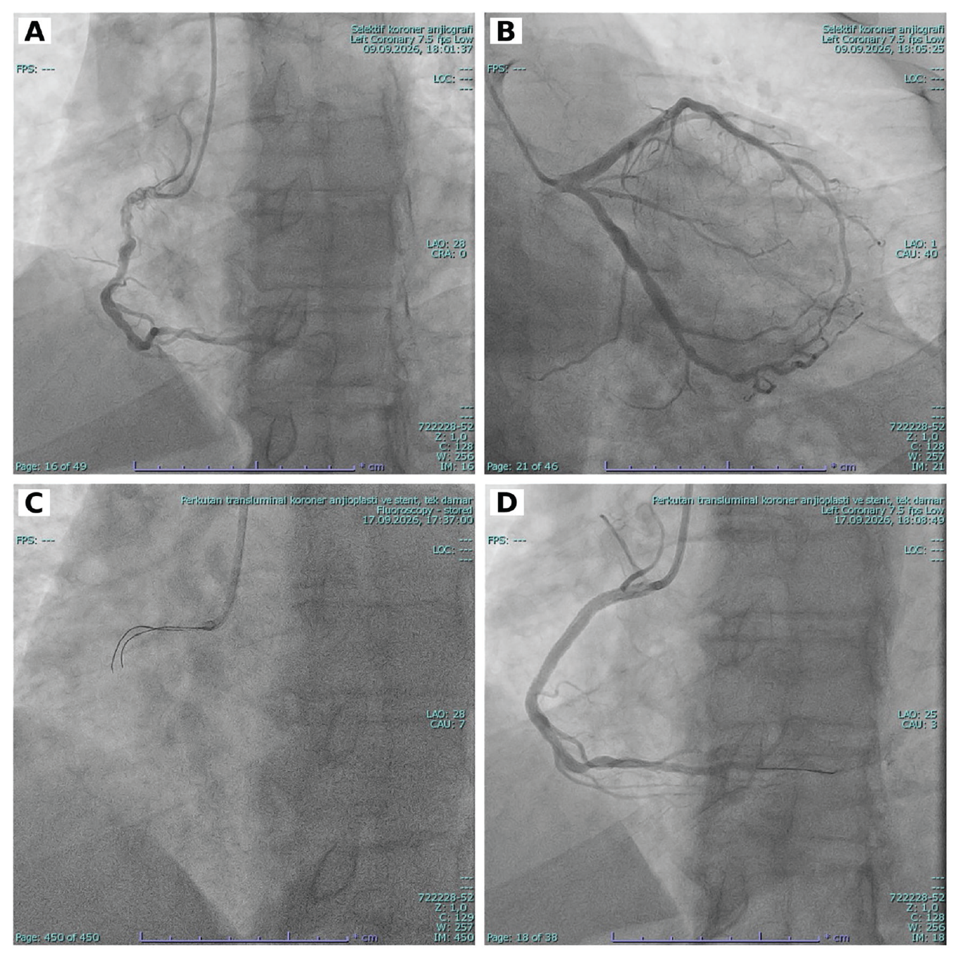
The circumflex lesion was treated with a 3.5×18-mm drug-eluting stent. Given the unusual morphology of the RCA lesion, an intravascular ultrasound (IVUS)-guided intervention was planned. Several guidewires, including Fielder and Choice wires, failed to cross the RCA lesion despite the use of microcatheter support (). Because the angiographic appearance suggested the presence of small, tortuous channels, a SUOH 03 wire was selected because its soft, flexible tip was considered suitable for navigating one of these channels. The wire was successfully advanced into the distal true lumen. Preintervention IVUS revealed multiple small lumens within a 40.1-mm woven-like segment, with a minimum lumen area of 1.94 mm2. The proximal and distal reference vessel diameters, measured at the external elastic membrane, were 4.45-4.54 mm and 4.46-4.73 mm, respectively (). Following sequential balloon predilation, a 3.5×48-mm drug-eluting stent was implanted and postdilated with a 4.0×18-mm non-compliant balloon (; ). Postintervention IVUS confirmed adequate stent expansion and apposition, with a minimum stent area of 11.13 mm2 ().
The patient was discharged one day after the procedure without in-hospital complications. On postprocedural day 15, he presented to the emergency department with chest pain. Troponin testing was negative, and echocardiography revealed no new abnormalities. An exercise stress test performed on the same day showed no evidence of inducible ischemia. The patient achieved an exercise capacity of 9.6 METs without experiencing chest pain during testing. No further episodes of chest pain were reported during the remainder of the 3-month follow-up.
This case illustrates the technical challenges of crossing a severely stenotic coronary segment with a woven-like angiographic appearance. Angiography alone cannot reliably distinguish a woven coronary artery from chronic dissection or recanalized thrombus., “Intravascular imaging can provide additional information about the intraluminal morphology of woven-like lesions., In the present case, preintervention IVUS findings supported the morphological assessment of the lesion; however, the angiographic appearance alone was insufficient to establish a definitive etiology.
Footnotes
Informed Consent: Written informed consent was obtained from the patient for publication of this case report and the accompanying images and videos.
Authorship Contributions: Surgical and Medical Practices: A.K., Concept: A.K, Design: M.T.Ö., Data Collection or Processing: M.T.Ö., Analysis or Interpretation: M.T.Ö., Literature Search: M.T.Ö., A.K., Writing: M.T.Ö.
Financial Disclosure: The authors declared that this study received no financial support.
Conflict of Interest: No conflict of interest was declared by the authors.
Declaration on the use of Artificial Intelligence (AI): AI-assisted technology was used to help edit the manuscript language and organize the revisions. The authors reviewed and take responsibility for the final content.
References
Yorifuji H, Shutta R, Egami Y, Nishino M, Tanouchi J. Woven coronary artery anomaly: optical coherence tomography versus intravascular ultrasound. JACC Case Rep. 2020;2:1698-1699.
Wen W, Liu H, Li J, Zhang Q. Woven-like change following intracoronary thrombosis recanalization: a case report. BMC Cardiovasc Disord. 2019;19:317.
Liu J, Hau WK, Li Y, Huang X. Application of a hybrid IVUS-OCT imaging system in woven-like coronary arteries. JACC Case Rep. 2025;30:105785.
Supplementary Materials
Video 1. Pre-intervention coronary angiography demonstrating the woven-like appearance of the proximal right coronary artery lesion
DownloadVideo 2. Pre-intervention intravascular ultrasound demonstrating multiple small lumens within the woven-like segment of the right coronary artery
DownloadVideo 3. Final angiographic appearance of the right coronary artery after drug-eluting stent implantation and post-dilation
DownloadVideo 4. Post-intervention intravascular ultrasound demonstrating final stent expansion and apposition, with a minimum stent area of 11.13 mm²
Download